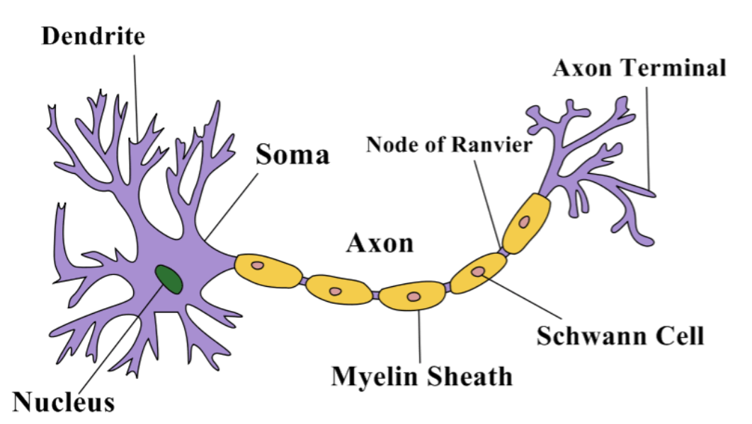
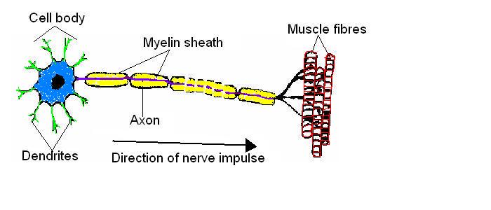
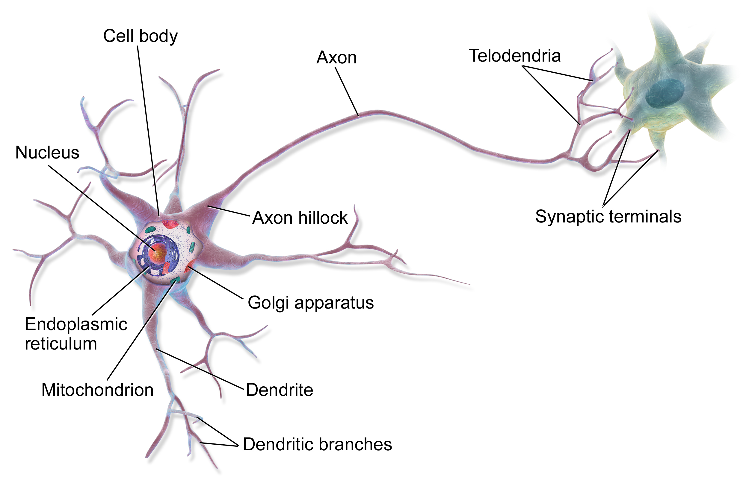
41 sketch and label a single motor neuron
Peripheral nerves: Histology and clinical aspects | Kenhub A single motor neuron may innervate several to several hundred muscle cells; collectively the unit is referred to as a motor end plate. The telodendria are rich in synaptic vesicles and mitochondria. The synaptic vesicles contain acetylcholine (ACh), which is released from the telodendria under the influence of the action potential. Neuron action potentials: The creation of a brain signal - Khan Academy When the brain gets really excited, it fires off a lot of signals. How quickly these signals fire tells us how strong the original stimulus is - the stronger the signal, the higher the frequency of action potentials. There is a maximum frequency at which a single neuron can send action potentials, and this is determined by its refractory periods.
Motor unit: definition and diagram | GetBodySmart 2. A motor unit is the term applied to a single motor neuron and all of the muscle fibers that it stimulates. Here's how to learn all muscles with quizzes and labeled diagrams. 1. 2. When a motor neuron fires, all the muscle fibers in the motor unit contract at once. The size of a motor unit varies from just a few fibers in the eye muscles ...

Sketch and label a single motor neuron
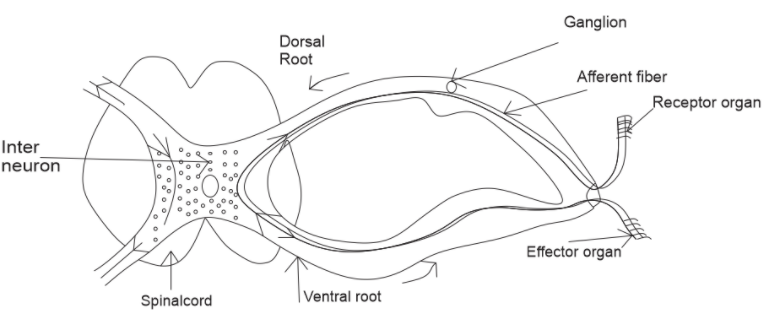
Sketch and label a single sensory neuron cell body.... Sketch and label a single sensory neuron cell body. ScienceBiologyAnimal Physiology Answer Step #1 of 2 A sensory neuron detects and responds to external stimuli. The sensory neurons receive signals with the help of receptors which are part of the peripheral nervous system. The received signals are converted into electrical impulses. Step #2 of 2 6.5.2 Draw and label a diagram of the structure of a motor neuron 6.5.2 Draw and label a diagram of the structure of a motor neuron 36,484 views Dec 1, 2013 127 Dislike Share Save Stephanie Castle 20.5K subscribers 4.9M views 12 years ago Aasoka 200K views... Schwann Cell Anatomy - Human Anatomy - GUWS Medical Sketch a single neuroglial cell in the space provided in Part C of the laboratory report. 11. Obtain a prepared microscope slide of a nerve. Locate the cross section of the nerve, and note the many round nerve fibers inside. Nerve fiber is a Figure 25.1 Label this diagram of a motor neuron. Figure 25.1 Label this diagram of a motor neuron.
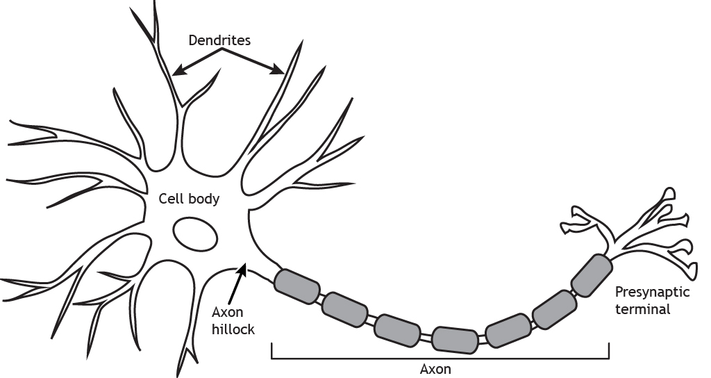
Sketch and label a single motor neuron. Label Parts of a Neuron Diagram | Quizlet Label Parts of a Neuron 4.2 (13 reviews) + − Flashcards Learn Test Match Created by cottonje Terms in this set (14) Term Dendrites Definition receives impulses from other nerve cells + 1 more side axon hillock The cell body...the part of the cell that houses the nucleus and keeps the entire cell alive and functioning Myelin Sheath Motor Neuron: Function, Types, and Structure - SimplyPsychology.org The structure of a motor neuron can be categorized into three components: the soma, the axon, and the dendrites. The soma is the cell body where the nucleus lies, which controls the cells and is also where proteins are produced to maintain the functioning of the neuron. The dendrites are the branch-like structures found at the ends of the neuron. How to draw a Motor Neuron - YouTube Hello friends in this video I will show you how to draw a neuron.Drawing motor neuron diagram or drawing neuron cell body is well shown in the video. The Neu... 35 Sketch And Label A Single Motor Neuron 652 draw and label a diagram of the structure of a motor neuron stephanie castle. Show where the concentrations of na and k are highest...
AP Biology Chapter 48 Flashcards | Quizlet This sketch shows two neurons. Label the following elements of this figure: cell body, dendrites, axon, synapse, presynaptic cell, postsynaptic cell, synaptic vesicles, synaptic terminal, and neurotransmitter. cell body: most organelles, including nucleus, are here Neurons (With Diagram) - Biology Discussion A neuron is a structural and functional unit of the neural tissue and hence the neural system. Certain neurons may almost equal the length of body itself. Thus neurons with longer processes (projections) are the longest cells in the body. Human neural system has about 100 billion neurons. Majority of the neurons occur in the brain. MOTOR UNITS AND MUSCLE TWITCHES - Brigham Young University-Idaho The motor neurons that innervate skeletal muscle fibers are called alpha motor neurons. As the alpha motor neuron enters a muscle, it divides into several branches, each innervating a muscle fiber (note this in the image above). One alpha motor neuron along with all of the muscle fibers it innervates is a motor unit . A Labelled Diagram Of Neuron with Detailed Explanations - BYJUS Diagram Of Neuron with Labels Here is the description of human neuron along with the diagram of the neuron and their parts. The neuron is a specialized and individual cell, which is also known as the nerve cell. A group of neurons forms a nerve.
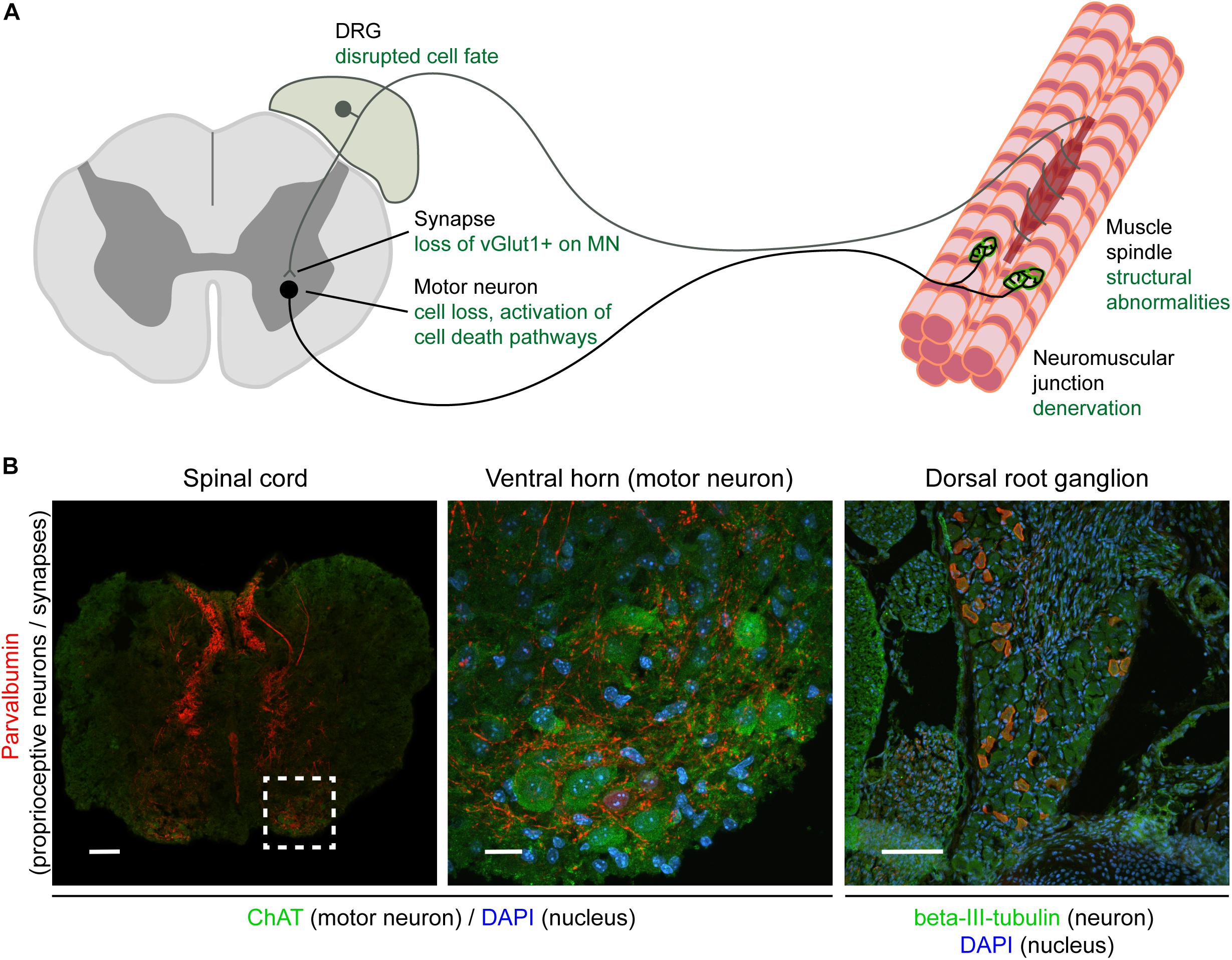
Sketch a neuron, Label the axon(s), dendrite(s), synaptic knob,... Sketch a neuron, Label the axon (s), dendrite (s), synaptic knob, Schwann cell, and a node of Ranvier. Indicate with an arrow which direction (s) nerve impulses flow. What would happen if there were more than one axon? Or what if there was only one dendrite? Sketch an action potential. Labeled Neuron Diagram - Science Trends Motor neurons are part of the central nervous system (CNS) and communicate signals from the spinal cord to the parts of the body to control their motion. For example, motor neurons send signals to the muscles in your arms causing them to contract. Motor neurons send electrical signals to your intestines so they move and churn food. Motor Neuron - The Definitive Guide | Biology Dictionary A motor neuron is a cell of the central nervous system. Motor neurons transmit signals to muscle cells or glands to control their functional output. When these cells are damaged in some way, motor neuron disease can arise. This is characterized by muscle wasting (atrophy) and loss of motor function. Motor Neuron Overview Schwann Cell Anatomy - Human Anatomy - GUWS Medical Sketch a single neuroglial cell in the space provided in Part C of the laboratory report. 11. Obtain a prepared microscope slide of a nerve. Locate the cross section of the nerve, and note the many round nerve fibers inside. Nerve fiber is a Figure 25.1 Label this diagram of a motor neuron. Figure 25.1 Label this diagram of a motor neuron.
6.5.2 Draw and label a diagram of the structure of a motor neuron 6.5.2 Draw and label a diagram of the structure of a motor neuron 36,484 views Dec 1, 2013 127 Dislike Share Save Stephanie Castle 20.5K subscribers 4.9M views 12 years ago Aasoka 200K views...
Sketch and label a single sensory neuron cell body.... Sketch and label a single sensory neuron cell body. ScienceBiologyAnimal Physiology Answer Step #1 of 2 A sensory neuron detects and responds to external stimuli. The sensory neurons receive signals with the help of receptors which are part of the peripheral nervous system. The received signals are converted into electrical impulses. Step #2 of 2

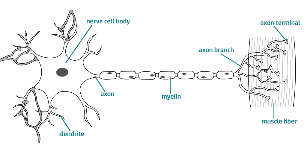








![2: Structure of a motor neuron [12]. | Download Scientific ...](https://www.researchgate.net/publication/308369018/figure/fig2/AS:654363412402179@1533023803638/Structure-of-a-motor-neuron-12.png)




Post a Comment for "41 sketch and label a single motor neuron"