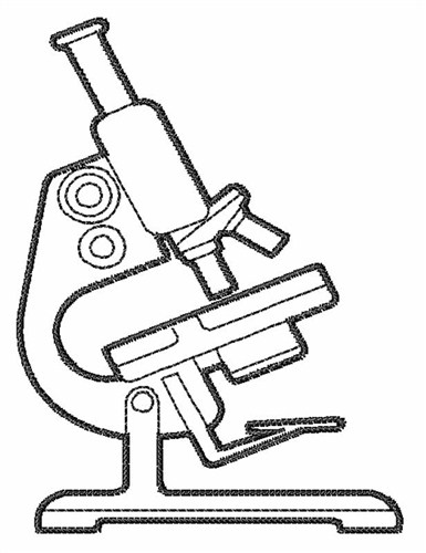
45 microscope drawing with labels
Pathology Outlines - Tuberculosis Definition / general. Due to Mycobacteria tuberculosis. In developing world, M. bovis causes oropharyngeal and intestinal TB. Very prevalent, approximately 1.7 billion people are infected with 9 million new cases a year with an estimated 1.6 million deaths. In the United States, there are 11,000 new cases of active TB each year. Gram Stain Technique - Amrita Vishwa Vidyapeetham Wipe the glass slide with spirit and wave the slide over the Bunsen burner to remove any unwanted microorganisms in the slide. Label one side of the glass slide with 1. Your initials 2. The date While flaming the inoculation loop be sure that each segment of metal glows orange/red-hot before you move the next segment into the flame.
lili ; LF moots on Twitter: "so our zoology lab prof made us draw 30 ... so our zoology lab prof made us draw 30 microscope slides of diff tissues with their labels and functions only to tell us to not submit it to him and use it as a reviewer instead 1:50 PM · Oct 7, 2022 · Twitter Web App
Microscope drawing with labels
Microscope: Types of Microscope, Parts, Uses, Diagram - Embibe A compound microscope is defined as a microscope with a high resolution. It uses two sets of lenses, providing a \ (2\)-dimensional image of the sample. The term compound refers to the usage of more than one lens in the microscope. Also, the compound microscope is one of the types of optical microscopes. Earthworm Dissection Draw a labeled illustration of and take notes on everything you view. External View, Four Pairs of Setae Labeled Setae Feel the bristly setae ( seta = bristle) on each segment, along the sides and bottom. Note that there are four pairs. DNA - Wikipedia Deoxyribonucleic acid (/ d iː ˈ ɒ k s ɪ ˌ r aɪ b oʊ nj uː ˌ k l iː ɪ k,-ˌ k l eɪ-/ (); DNA) is a polymer composed of two polynucleotide chains that coil around each other to form a double helix carrying genetic instructions for the development, functioning, growth and reproduction of all known organisms and many viruses.DNA and ribonucleic acid (RNA) are nucleic acids.
Microscope drawing with labels. Library Home / LibGuides: Chemistry I & II: The Lab Report Data: Use charts, graphs, and tables to organize your data in clearly labeled sections. Number and title every table. Have a caption for every chart or graph. An example of the calculations or formulas should be provided. Any outside information must be referenced properly. Do not include any interpretation or opinions. Metaphase - Genome.gov During metaphase, the nucleus dissolves and the cell's chromosomes condense and move together, aligning in the center of the dividing cell. At this stage, the chromosomes are distinguishable when viewed through a microscope. Metaphase chromosomes are used in karyotyping, a laboratory technique for identifying chromosomal abnormalities. Narration Cell Division: Mitosis and Meiosis - Owlcation When examined on them, try to use labelled diagrams and tables to summarise the key information. Also ensure that all key terms are used appropriately, as this can save you time and energy. The Stages of Mitosis In this slide you can see the upper cell in Prophase, and the lower cell well into Anaphase 1 / 3 Mitosis Stages THE TRAJECTORY OF U.S. FOREIGN POLICY - The Greanville Post JOHN RACHEL—This regime of perpetual war and global domination is the work of madmen, power-drunk sociopaths who've grabbed and now maintain absolute control of our foreign policy. They are empire-obsessed megalomaniacs who've seized the initiative and are the architects of the Great Imperial Project — the U.S. as absolute imperial master of the Earth. They have without any consent by ...
Medicine & Health | UNSW Sydney Research & impact. UNSW Medicine & Health is world renowned for its research and impact addressing issues of health disparities and improving lives through our key research themes of Cancer; Infectious Disease, Immunity & Inflammation; Neuroscience, Mental Health & Addiction; Cardiac, Vascular & Metabolic Medicine and Health Systems. X-ray crystallography - Wikipedia X-ray crystallography is the experimental science determining the atomic and molecular structure of a crystal, in which the crystalline structure causes a beam of incident X-rays to diffract into many specific directions. By measuring the angles and intensities of these diffracted beams, a crystallographer can produce a three-dimensional picture of the density of electrons within the crystal. Mitochondria - Genome.gov Mitochondria are membrane-bound cell organelles (mitochondrion, singular) that generate most of the chemical energy needed to power the cell's biochemical reactions. Chemical energy produced by the mitochondria is stored in a small molecule called adenosine triphosphate (ATP). Mitochondria contain their own small chromosomes. Colony Morphology of Bacteria - Microbe Online The six most common elevations of bacterial colonies are flat, raised, umbonate (having a knobby protuberance), crateriform, convex, and pulvinate (cushion-shaped). The margin of the bacterial colony: The margin or edge of a colony may be a vital characteristic in identifying organisms. Examples are entire (smooth), irregular, undulate (wavy),
The latest in Machine Learning | Papers With Code VToonify: Controllable High-Resolution Portrait Video Style Transfer. williamyang1991/vtoonify • • 22 Sep 2022 Although a series of successful portrait image toonification models built upon the powerful StyleGAN have been proposed, these image-oriented methods have obvious limitations when applied to videos, such as the fixed frame size, the requirement of face alignment, missing non ... DP Biology: Calculating Magnification and Size Activity 1 Calculating magnification of an image using it's scale bar The three images below (click the eye to reveal) show a worked example of how to calculate sizes of cells organelles from electron micrographs step by step. Follow these steps carefully then complete the calculations on the worksheet. X-Ray Generation Notes - University of Oklahoma Two cartoons of an X-ray tube. Drawing a) shows the line and spot focus patterns of a typical sealed tube. Drawing b) shows the take-off angle of a tube. The generation of X rays is very inefficient. In addition to white radiation and characteristic lines, laboratory sources also produce Auger electrons and photo-electrons. TMZ Sports | Latest News, Videos & Photos TMZ Sports has learned the Packers organization reached out to celeb artist Joe Castro last month -- in order to have custom spikes made for their 25-year-old cornerback for their big game against ...
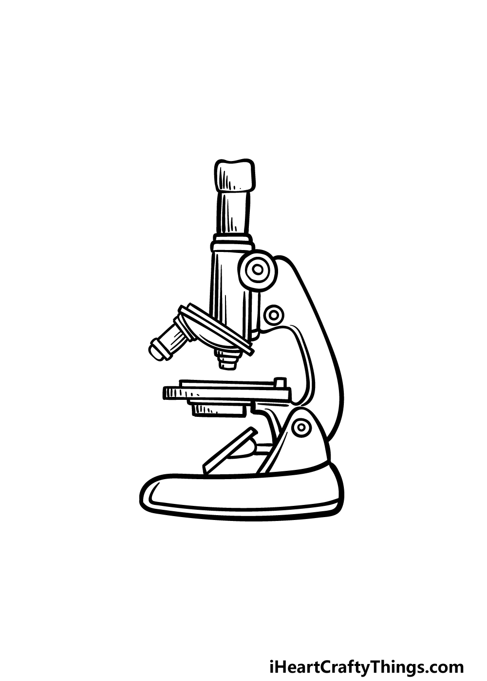
Microscope Drawing - How To Draw A Microscope Step By Step
KOH Mount: Principle, Procedure, Results, Uses - Microbe Online Weigh 10 g potassium hydroxide (KOH) pellets. Transfer the chemical to a screw-cap bottle. Add 50 ml distilled water, and mix until the chemical is completely dissolved. Add remaining distilled water and make the volume 100 ml. Label the bottle and mark it as corrosive. Store it at room temperature. The reagent is stable for up to 2 years.

Simple doodles, Microscope parts, Biology labs
13 Best Image Annotation Tools of 2022 [Reviewed] - V7Labs Supports most unique file types (ultra-high-resolution, multi-spectral, microscopy formats, PDF) Labelbox Labelbox is a training data platform built from three core layers that facilitate the entire process from labeling and collaboration to iteration. It was created in 2018 and has quickly become one of the most popular data labeling tools.

label microscope diagram | Charts | Microscope, Anatomy bones ...
Form 4 Biology Pp3 Exams (Questions, Confidential & Answers) Draw and label specimen labeled D2 (3marks) Giving a reason and state the agent of dispersal of the specimen (6marks) Specimen : Agent of dispersal ... 1.You are provided with the photomicrograph of an onion outer epidermis as seen under light microscope . a) On the photograph, name parts labelled A, C, and D (3marks) A chloroplast ; C cell ...

How to draw Compound microscope | Microscope diagram easily Draw | SSC diagram
Programmable RNA sensing for cell monitoring and manipulation Binary vectors labeled cells shown in cortex (c1). c2, Magnified view of boxed region in c1. vGAT mRNAs were labeled by in-situ hybridization (c3). Co-labeling by mNeon and vGAT mRNA (c4). Arrows ...
Microscope- Simple-AND Compound-WITH- Label - BS in Education ...
Keep it or toss it? 'Best Before' labels cause confusion But ReFED estimates that 7% of U.S. food waste — or 4 million tons annually — is due to consumer confusion over "best before" labels. Date labels were widely adopted by manufacturers in ...

how to draw diagram of microscope | how to draw diagram of microscope step by step | microscope
Inspection Checklists - Sample Checklist for Manufacturing Facilities The examples outlined below do not list all the possible items for manufacturing facilities. The best checklist for your workplace is one that has been developed for your specific needs. Whatever the format of the checklist, provide space for the inspectors' signatures and the date. Inspectors:

Cell Drawing Microscope - Binocular Compound Microscope ...
AQA | Search Use a light microscope to observe, draw and label a selection of plant and animal cells. ... Topic 1 - Key concepts in biology . Topic 5 - Homeostasis and response.
Free Microscope Drawing, Download Free Microscope Drawing png ...
5 White Blood Cells Types and Their Functions - New Health Advisor They are very colorful when stained and looked at under the microscope, making them easy to identify. Function: Basophils have the ability to secrete anticoagulants and antibodies that have function against hypersensitivity reactions in the bloodstream. They act immediately as part of the immune system's action against foreign invaders.

Diagram Of A Microscope | Diagram, Microscope, Bullet journal ...
scheme work biology - Free KCPE Past Papers Introduction to light microscope. By the end of the lesson, the learner should be able to: Define a cell; Draw and label the light microscope; Description of a cell; Drawing and labeling the light microscope . Light microscope; Diagram of light microscope; Comprehensive secondary Biology students Bk. 1 page 17; Teachers bk. 1 pages 11-19; KLB ...

Labeling the Parts of the Microscope | Microscope activity ...
Motility Test (Theory) - Amrita Vishwa Vidyapeetham Virtual Lab Three methods are employed for motility determination depending on the pathogenic capability of the organisms. For nonpathogens, there are two slide techniques that one might use. For pathogens, tube method can be used. I) Slide methods for non-pathogens include 1. Wet Mount slide 2. Hanging Drop slide 1. Wet Mount slide
![How To Draw A Microscope Step by Step - [12 Easy Phase]](https://easydrawings.net/wp-content/uploads/2021/01/Overview-for-Microscope-drawing.jpg)
How To Draw A Microscope Step by Step - [12 Easy Phase]
Gram Staining Procedure | New Health Advisor Draw a circle under the slides using a marking pen designed for glassware. This will help to designate which area to prepare the smear in the following step. You can also label them with the organism's initials at the edge of each slide. Take care that the labels do not get in contact with the reagentsused forstaining. 3. Prepare the Smear
![Very Easy!!! How to draw microscope [with labels]](https://i.ytimg.com/vi/mtXYI7wO-qs/maxresdefault.jpg)
Very Easy!!! How to draw microscope [with labels]
DNA - Wikipedia Deoxyribonucleic acid (/ d iː ˈ ɒ k s ɪ ˌ r aɪ b oʊ nj uː ˌ k l iː ɪ k,-ˌ k l eɪ-/ (); DNA) is a polymer composed of two polynucleotide chains that coil around each other to form a double helix carrying genetic instructions for the development, functioning, growth and reproduction of all known organisms and many viruses.DNA and ribonucleic acid (RNA) are nucleic acids.
Free Microscope Drawing, Download Free Microscope Drawing png ...
Earthworm Dissection Draw a labeled illustration of and take notes on everything you view. External View, Four Pairs of Setae Labeled Setae Feel the bristly setae ( seta = bristle) on each segment, along the sides and bottom. Note that there are four pairs.
Free Microscope Drawing, Download Free Microscope Drawing png ...
Microscope: Types of Microscope, Parts, Uses, Diagram - Embibe A compound microscope is defined as a microscope with a high resolution. It uses two sets of lenses, providing a \ (2\)-dimensional image of the sample. The term compound refers to the usage of more than one lens in the microscope. Also, the compound microscope is one of the types of optical microscopes.

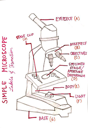
Simple Microscope - Parts, Functions, Diagram and Labelling ...

Compound Microscope Drawing With Parts and Functions

How to draw Microscope diagram for beginners - step by step
Free Microscope Drawing, Download Free Microscope Drawing png ...

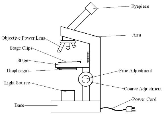
Diagram of a Compound Microscope
Free Microscope Drawing, Download Free Microscope Drawing png ...

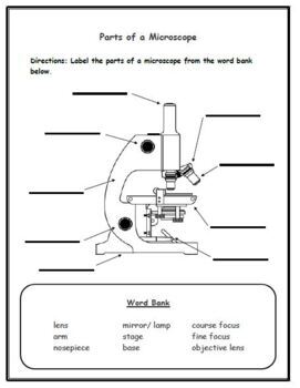
Parts of a Microscope - SmartSchool Systems

Microscope Drawing

Compound Microscope Parts – Labeled Diagram and their ...
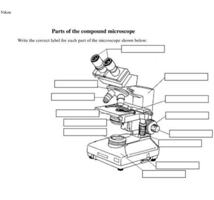
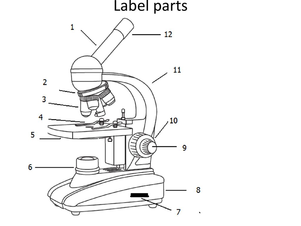
Solved Nikon Parts of the compound microscope Write the ...

Label the microscope — Science Learning Hub

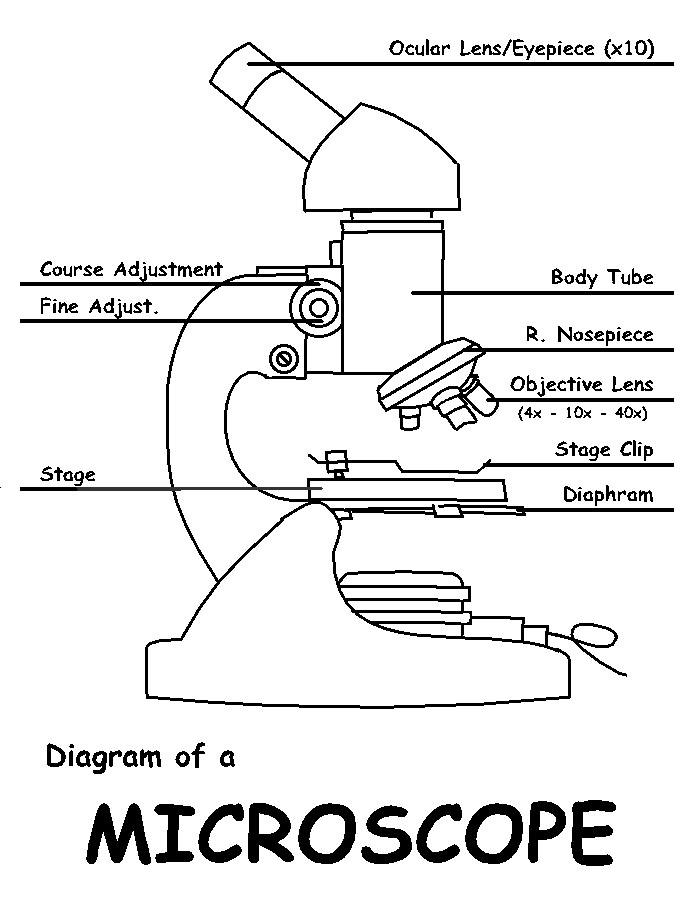
Compound Microscope Parts, Functions, and Labeled Diagram ...

A Study of the Microscope and its Functions With a Labeled ...

Compound Microscope Parts – Labeled Diagram and their ...

How to Draw a Microscope - VERY EASY

Dissecting Stereo Microscope Parts and Functions
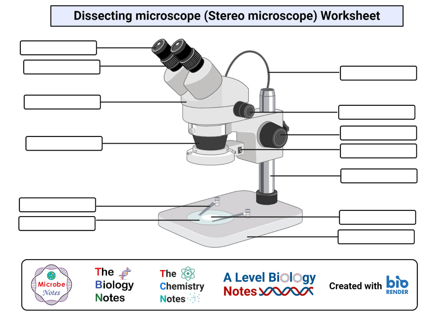
Parts of a microscope with functions and labeled diagram

Free Microscope Drawing, Download Free Microscope Drawing png ...

How to Draw a Microscope - Really Easy Drawing Tutorial

label the parts of the compound microscope - Brainly.ph

Free Microscope Drawing, Download Free Microscope Drawing png ...
Collection Of Free Microscopes Drawing Label Clipart ...

Simple Microscope- Definition, Principle, Magnification ...

Cytology. Cytology. radiation used to illuminate the specimen ...
Free Microscope Drawing, Download Free Microscope Drawing png ...

How to draw compound of Microscope easily - step by step
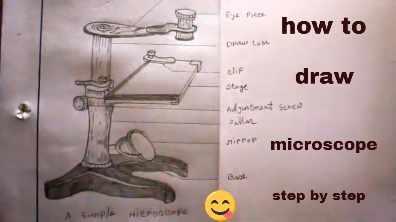
How TO Draw simple microscope step by step/simple microscope drawing/for science project
Label the Microscope Parts for Elementary School Students
Free Microscope Drawing, Download Free Microscope Drawing png ...

Microscope Diagram Labeled, Unlabeled and Blank | Parts of a ...
Compound Microscope Review - ppt download
Post a Comment for "45 microscope drawing with labels"