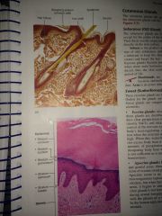
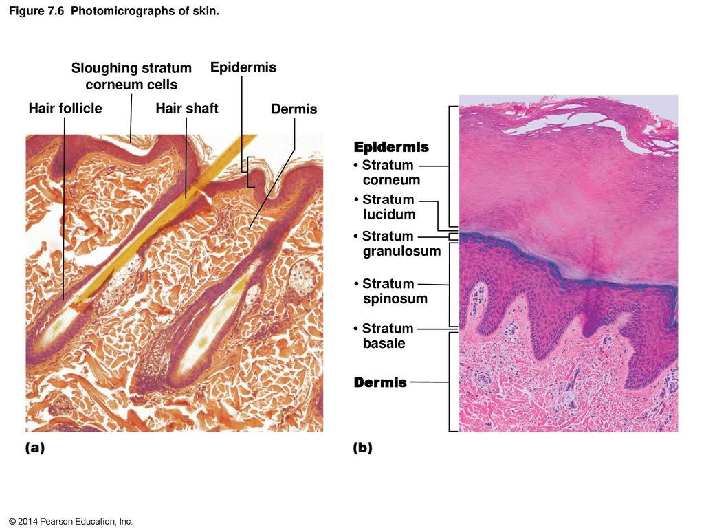
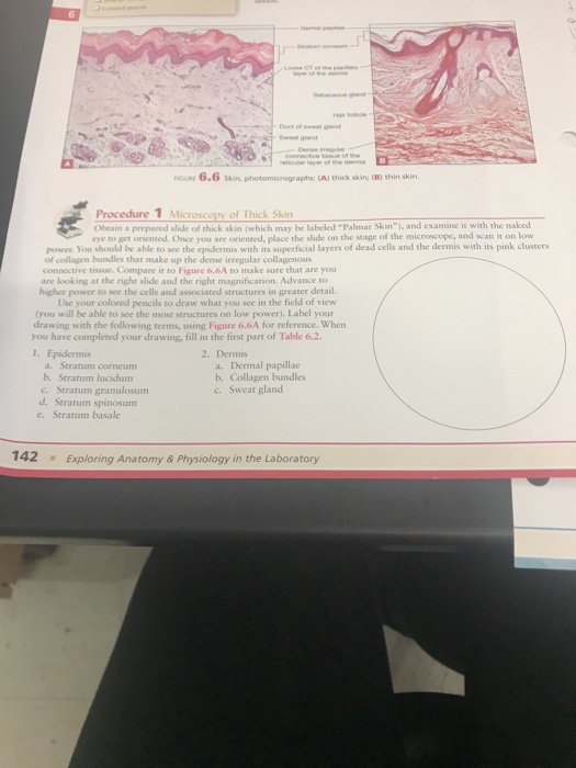
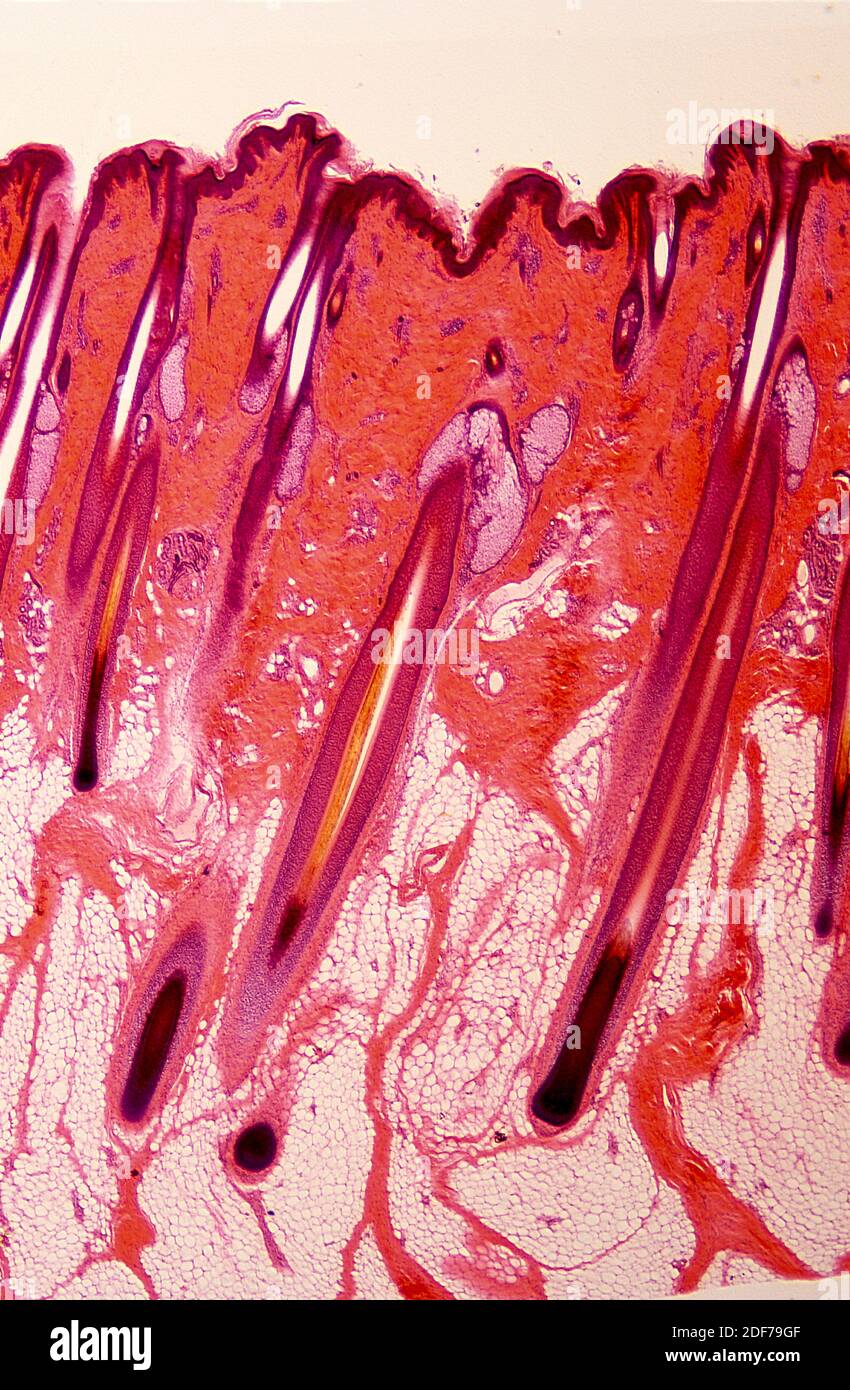
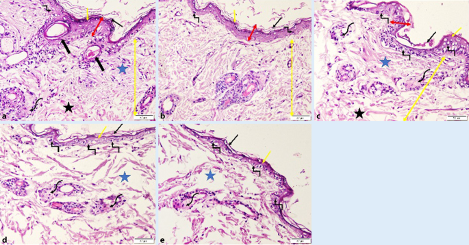
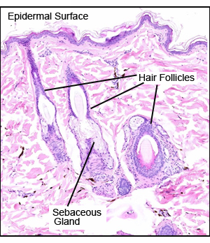
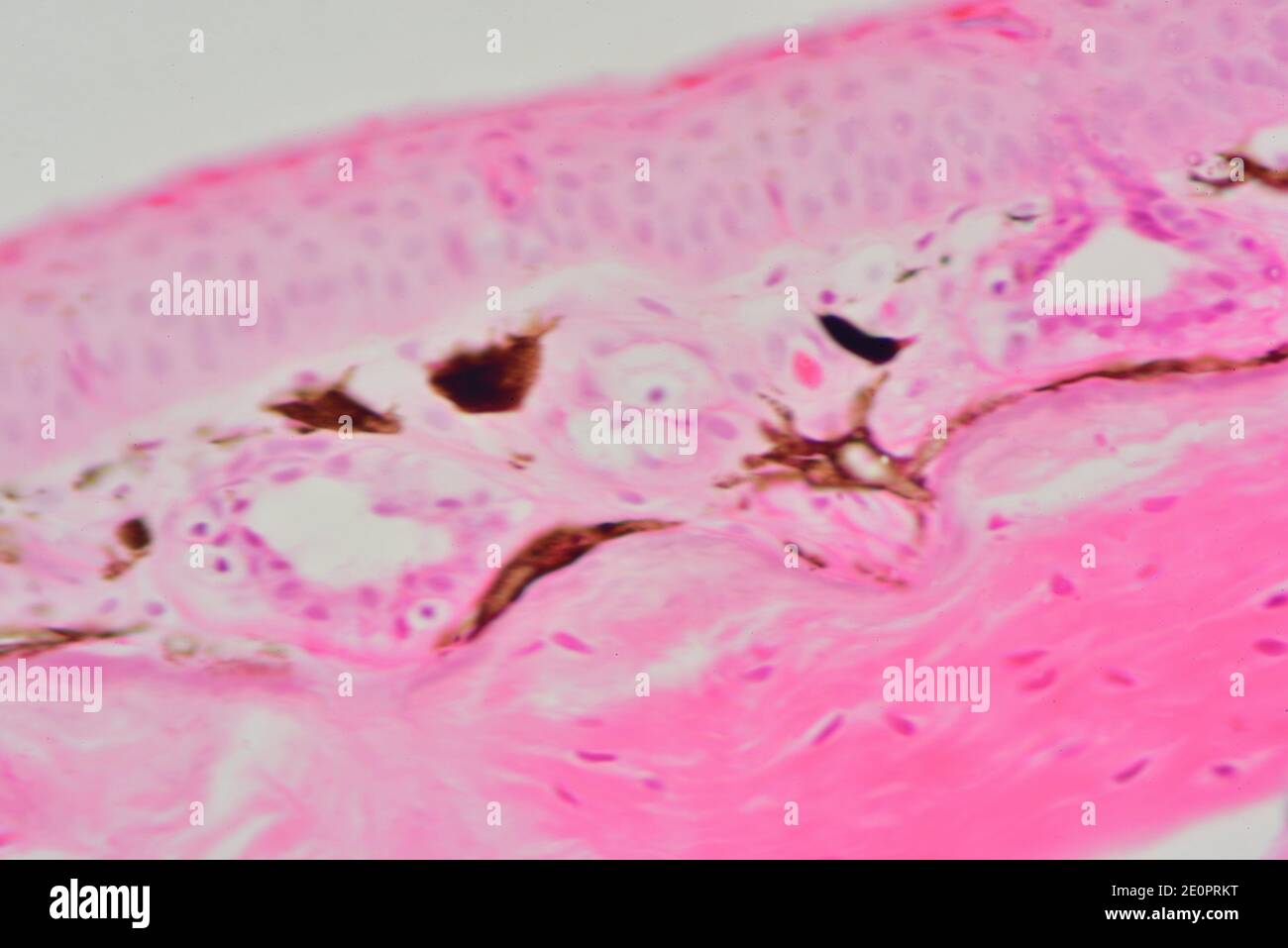
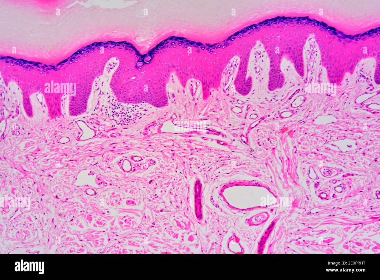
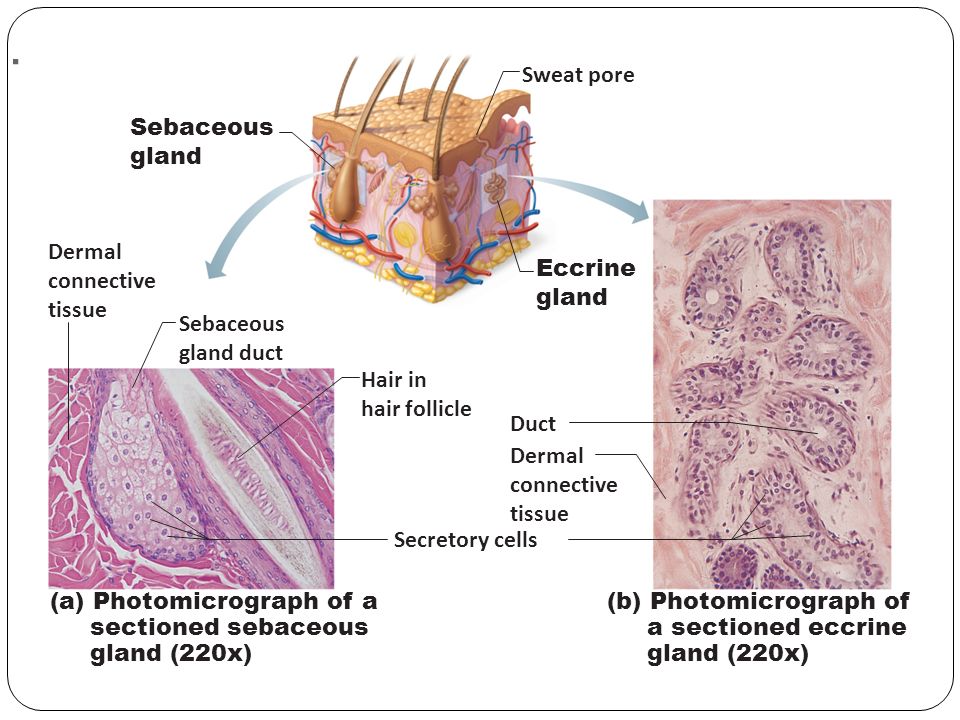
39 photomicrograph of thin skin labeled
rsscience.com › hair-under-a-microscopeHair Under a Microscope - Rs' Science The fur is a thick growth of hair that covers the skin of many mammals. It consists of a combination of longer guard hair on top and shorter fleece hair (also known as underfur or down hair) beneath. The guard hair keeps moisture from reaching the skin; the underfur acts as an insulating blanket that keeps the animal warm. Thermal insulation Label The Photomicrograph Of Thin Skin / Skin Cross Section High ... Some labeled features may be referred to once, more than once, or not at all. Stratum corneum stratum granulosum stratum spinosum stratum basale dermis epidermis. Label the photomicrograph of thin skin 1 answer below ». This is a photomicrograph of thin skin. The ducts are lined by stratified (2 layers) cuboidal epithelium. Label the ...
en.wikipedia.org › wiki › KaryotypeKaryotype - Wikipedia Cutting up a photomicrograph and arranging the result into an indisputable karyogram. The work took place in 1955, and was published in 1956. The karyotype of humans includes only 46 chromosomes. The other great apes have 48 chromosomes. Human chromosome 2 is now known to be a result of an end-to-end fusion of two ancestral ape chromosomes.

Photomicrograph of thin skin labeled
Label The Photomicrograph Of Thick Skin Quizlet : Solved Label The ... Label the photomicrograph of thin skin. Learn vocabulary, terms, and more with flashcards, games, and other study tools. In the diagram of skin shown below, which labeled structure generates fingerprints? But studies show that people who solicit and accept feedback are more effective leaders and more successful at work. Part a is a micrograph ... Anatomy and Physiology Homework Chapter 6 Flashcards | Quizlet Label the photomicrograph of thin skin.-Duct of sebaceous gland-Epidermis-Hair-Sebaceous gland-Dermis-Hair Follicle-Epidermis-Hair-Duct of sebaceous gland-Sebaceous gland-Hair Follicle-Dermis Explanation: Thin skin is located throughout the body. Refer to APR 3.0 for further information. quizlet.com › 431484880 › chapter-5-flash-cardsChapter 5 Flashcards | Quizlet Which of the following are characteristics of thick skin? Select all that apply. a) Found in the palms, soles of the feet and fingertips. b) Does not contain hair follicles. c) Contains more sweat glands than thin skin. d) Stratum corneum has fewer layers in thick than thin. e) Lacking stratum lucidum
Photomicrograph of thin skin labeled. Photomicrograph Of Thick Skin Labeled : Jaypeedigital Ebook Reader Label the photomicrograph of thick skin. Stratum corneum stratum basale stratum granulosum stratum lucidum epidermis dermis stratum . Learn vocabulary, terms, and more with flashcards, games, and other study tools. Thick skin has a thinner dermis than thin skin, and does not contain hairs, sebaceous glands, . Learn more about skin discoloration ... Label The Photomicrograph Of Thin Skin. - Skin Model 1 - YouTube Hair sebaceous gland dermis hair follicle epidermis duct of sebaceous. Label the photomicrograph of thin skin. This is a photomicrograph of thin skin. Learn more about thin skin treatment at howstuffworks. It has a fifth layer, called the stratum lucidum, located between the stratum corneum and the stratum granulosum (figure 2). Thin skin ... › 43095131 › Fundamentals_ofFundamentals of Analytical Chemistry- 9th Edition - Academia.edu Enter the email address you signed up with and we'll email you a reset link. Sebaceous Gland Label The Photomicrograph Of Thin Skin - Integumentary ... Label the photomicrograph of thin skin. (b) a photomicrograph of he section of thin skin tissue from burnt . Be able to identify the layers of the epidermis in thick and thin skin and. The ducts are lined by stratified (2 layers) cuboidal epithelium. Name the 4 layers of thin skin in both the cartoon and the photomicrograph.
› articles › s41598/021/97778-3A Tunguska sized airburst destroyed Tall el-Hammam a Middle ... Sep 20, 2021 · We present evidence that in ~ 1650 BCE (~ 3600 years ago), a cosmic airburst destroyed Tall el-Hammam, a Middle-Bronze-Age city in the southern Jordan Valley northeast of the Dead Sea. Label The Photomicrograph Of Thin Skin And Its Accessory Structures ... The skin and its accessory structures make up the integumentary system, which provides the. Part a is a micrograph showing a cross section of thin skin. Accessory structures of the skin include hair, nails, sweat glands, and sebaceous glands. The nail bed is a . Name the 4 layers of thin skin in both the cartoon and the photomicrograph. Label The Photomicrograph Of Thick Skin. - Exercise 4 Quiz Flashcards ... 1 answer to label the photomicrograph of thin skin. The epidermis, made of closely packed epithelial cells, and the dermis, made of dense, irregular connective tissue . Epidermis Of Thick Skin from eugraph.com The skin is composed of two main layers: Thick skin showing epithelial detail. Practice labeling the layers of the skin. Solved Label the photomicrograph of thin skin. Dermis Duct - Chegg Question: Label the photomicrograph of thin skin. Dermis Duct of sebaceous gland Hair Follicle Sebaceous gland Hair Epidermis This problem has been solved! See the answer Show transcribed image text Expert Answer 100% (35 ratings) A … View the full answer Transcribed image text: Label the photomicrograph of thin skin.
Photomicrograph of Thin Skin Quiz - PurposeGames.com This is an online quiz called Photomicrograph of Thin Skin. There is a printable worksheet available for download here so you can take the quiz with pen and paper. Your Skills & Rank. Total Points. 0. Get started! Today's Rank--0. Today 's Points. One of us! Game Points. 5. Label The Photomicrograph Of Thick Skin - Faktor yang Label the photomicrograph of thick skin. 1 answer to label the photomicrograph of thin skin. The epidermis of thick skin has five layers: Hypodermis label the layers of the epidermis in thick skin in figure 7.2. A few layers of cells that are . Apocrine sweat gland label the photomicrograph in figure 7.4. Label the photomicrograph of thick skin. EOF › 28860759 › Material_Science_andMaterial Science and Engineering, seventh edition - Academia.edu Enter the email address you signed up with and we'll email you a reset link.
› 40521511 › Janeways_ImmunobiologyJaneway's Immunobiology - 9th Edition - Academia.edu Janeway's Immunobiology is a textbook for students studying immunology at the undergraduate, graduate, and medical school levels. As an introductory text, students will appreciate the book's clear writing and informative illustrations, while
Solved Thin skin histology HM 44 Label the photomicrograph - Chegg Science. Anatomy and Physiology. Anatomy and Physiology questions and answers. Thin skin histology HM 44 Label the photomicrograph of thin skin. points 01:04:56 Stratum granulosum eBook Stratum spinosum Dermis Epidermis Stratum corneum Stratum basale.
Label The Photomicrograph Of Thin Skin And Its Accessory Structures ... Photomicrograph Of Thin Skin Labeled - NaturalSkins from d2vlcm61l7u1fs.cloudfront.net Figure 7.2 the main structural features in epidermis of thin skin. Label the photomicrograph of the skin and its accessory structures. These originate embryologically from the epidermis and . Part a is a micrograph showing a cross section of thin skin. Label ...
Photomicrograph Of Thick Skin Labeled : Integument Sciencedirect Solved 21 Label The Photomicrograph Of Thick Skin Chegg Com from media.cheggcdn.com Cornified (keratinized) stratified squamous epithelium makes up the epidermis. 1 answer to label the photomicrograph of thin skin. Lucidum, present in thick skin, is not illustrated here. It has a fifth layer, called the stratum lucidum, located between the ...
Question : Question 31 points Label the photomicrograph of thin skin ... Question 31. A first grade teacher wishes to "shape" her student's writing of the alphabet. The teacher should: a, reward the child whenever the c... Question 31. A neurotransmitter that allows sodium ions to leak into a postsynaptic neuron causes: A) inhibitory postsynaptic damage to the myelin sheath C) excitatory postsynaptic...
photomicrographs of thin skin Flashcards | Quizlet Only $35.99/year photomicrographs of thin skin STUDY Flashcards Learn Write Spell Test PLAY Match Gravity Created by Madison_Tacquard Terms in this set (4) stratum corneum sebaceous gland hair follicle dense irregular CT of the reticular layer of the dermis Sets found in the same folder hair structure 8 terms Madison_Tacquard nail anatomy 11 terms
quizlet.com › 431484880 › chapter-5-flash-cardsChapter 5 Flashcards | Quizlet Which of the following are characteristics of thick skin? Select all that apply. a) Found in the palms, soles of the feet and fingertips. b) Does not contain hair follicles. c) Contains more sweat glands than thin skin. d) Stratum corneum has fewer layers in thick than thin. e) Lacking stratum lucidum
Anatomy and Physiology Homework Chapter 6 Flashcards | Quizlet Label the photomicrograph of thin skin.-Duct of sebaceous gland-Epidermis-Hair-Sebaceous gland-Dermis-Hair Follicle-Epidermis-Hair-Duct of sebaceous gland-Sebaceous gland-Hair Follicle-Dermis Explanation: Thin skin is located throughout the body. Refer to APR 3.0 for further information.
Label The Photomicrograph Of Thick Skin Quizlet : Solved Label The ... Label the photomicrograph of thin skin. Learn vocabulary, terms, and more with flashcards, games, and other study tools. In the diagram of skin shown below, which labeled structure generates fingerprints? But studies show that people who solicit and accept feedback are more effective leaders and more successful at work. Part a is a micrograph ...
:max_bytes(150000):strip_icc()/5324695-GettyImages-139812232-75c6744d0b2246fba58223c0eb784c73.jpg)














Post a Comment for "39 photomicrograph of thin skin labeled"